Gynaec and Obstetric Sonography
Gynaec and Obstetric Sonography
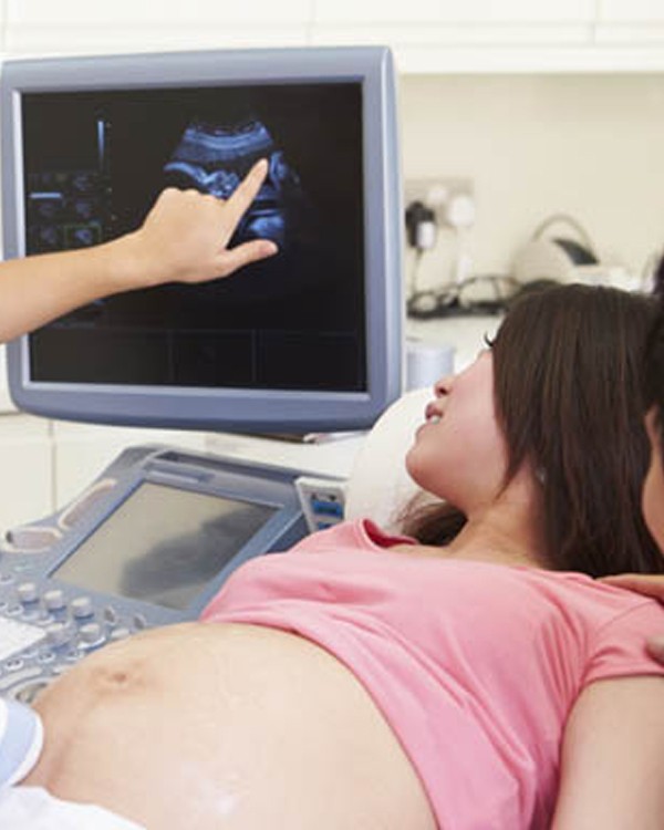
Gynaec and Obstetric Sonography, also known as ultrasound imaging in gynecology and obstetrics, has become an essential tool in modern medicine, particularly in the care and management of women’s reproductive health. Its roots are deeply tied to a pivotal moment in medical history that dates back to 1958. While many advancements in medicine evolve gradually and lack a definitive starting point, the use of ultrasound in obstetrics and gynecology began with remarkable clarity through a landmark publication in The Lancet by Ian Donald, John McVicar, and Tom Brown. Their paper, titled “The investigation of abdominal masses by pulsed ultrasound,” marked the beginning of a new era.
Although the title of the 1958 paper does not reflect its groundbreaking nature, its content was revolutionary. It introduced the world’s first ultrasound images of the human fetus and gynecological masses, captured using a compound contact scanner—the first practical ultrasound scanning machine. This advancement transformed what had previously been a theoretical or experimental concept into a real and highly useful clinical tool. The ability to see inside the womb non-invasively was nothing short of revolutionary and has since changed the face of prenatal and gynecological care forever.
From this foundation, gynaec and obstetric sonography rapidly developed. In obstetrics, ultrasound became indispensable for monitoring fetal development, assessing fetal growth, detecting congenital anomalies, and determining gestational age. It plays a vital role in managing high-risk pregnancies, identifying complications such as ectopic pregnancies, placenta previa, or multiple gestations, and guiding critical procedures like amniocentesis or chorionic villus sampling.
In gynecology, sonography provides detailed images of the uterus, ovaries, and surrounding pelvic structures. It is commonly used to evaluate abnormal uterine bleeding, fibroids, ovarian cysts, endometrial thickness, and uterine anomalies. Transvaginal ultrasound, a more recent advancement, offers even greater resolution and diagnostic accuracy for early pregnancy evaluation and detailed pelvic assessments.
The evolution of ultrasound technology has continued over the decades, with improvements such as real-time imaging, Doppler studies, 3D/4D imaging, and portable machines making sonography more accurate, accessible, and patient-friendly. 3D and 4D imaging, in particular, have allowed for more detailed visualization of the fetus, enhancing both diagnostic capabilities and the emotional experience for expectant parents.
Today, gynaec and obstetric sonography is a cornerstone of women’s healthcare and is routinely used in clinics, hospitals, and diagnostic centers across the globe. Its safety, non-invasiveness, and affordability make it an ideal imaging method for reproductive health.
In conclusion, the field of gynecological and obstetric sonography owes its origins to the trailblazing work of Donald, McVicar, and Brown in 1958. What started as an experimental procedure has grown into an indispensable clinical tool that supports the diagnosis, monitoring, and management of countless conditions related to pregnancy and female reproductive health. As technology continues to advance, so too will the capabilities of ultrasound in improving outcomes for women and their babies.